Slime mold illustrations for an Australian guidebook
By Dr. Stephen L. Stevenson
Colorful, tiny, spore-filled organisms like these may live on a dead log near you.
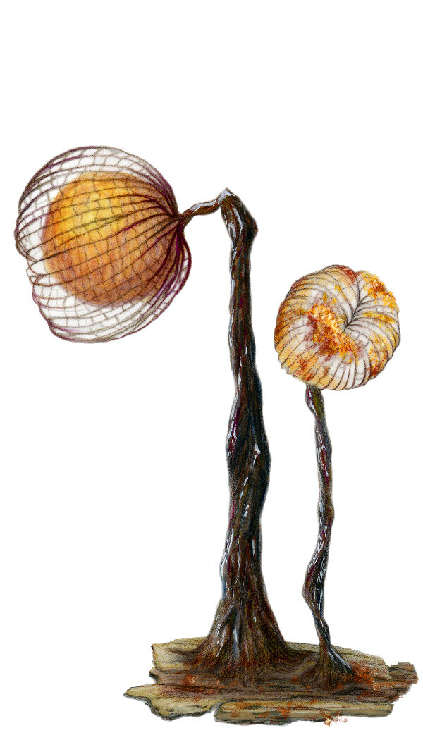
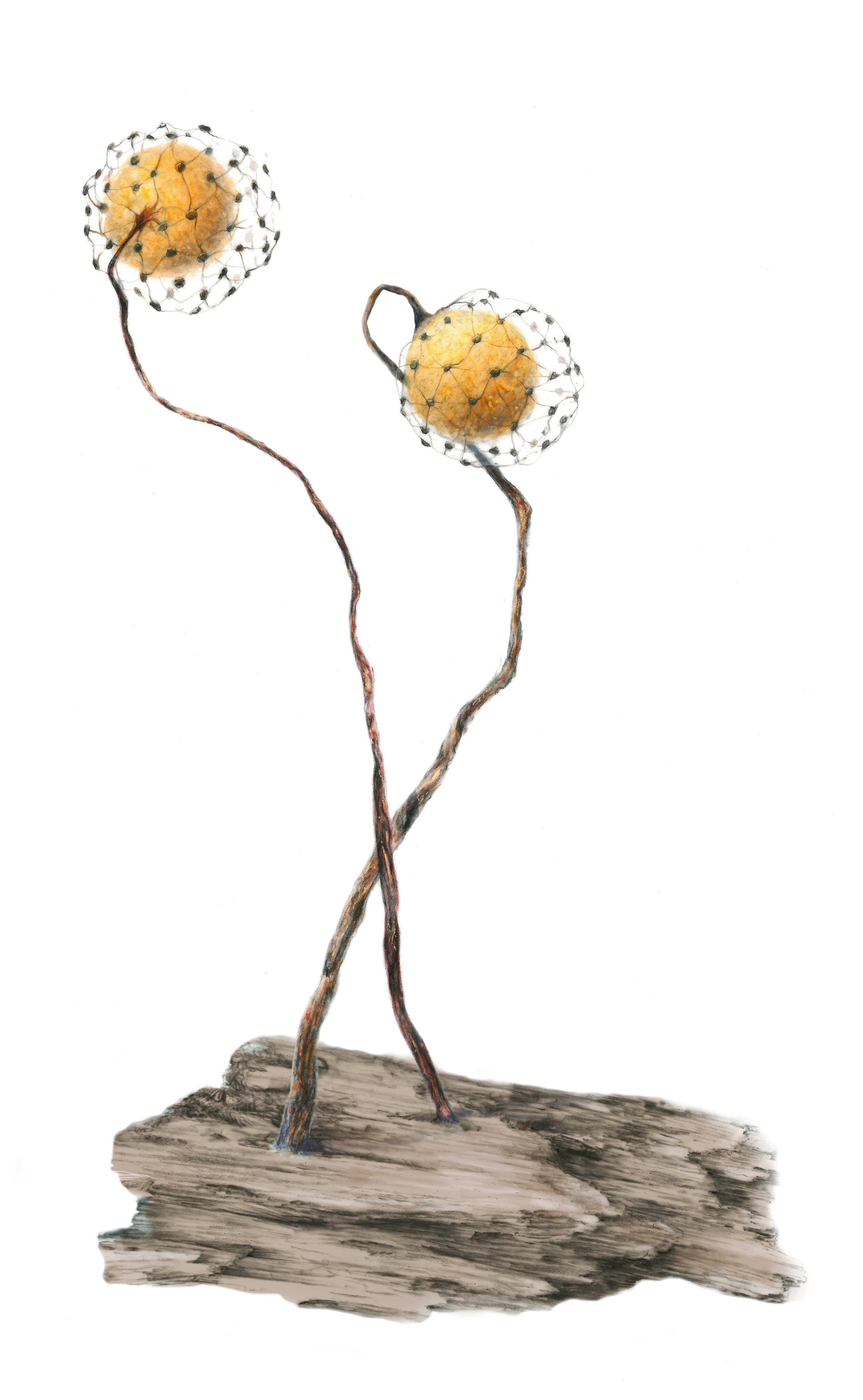
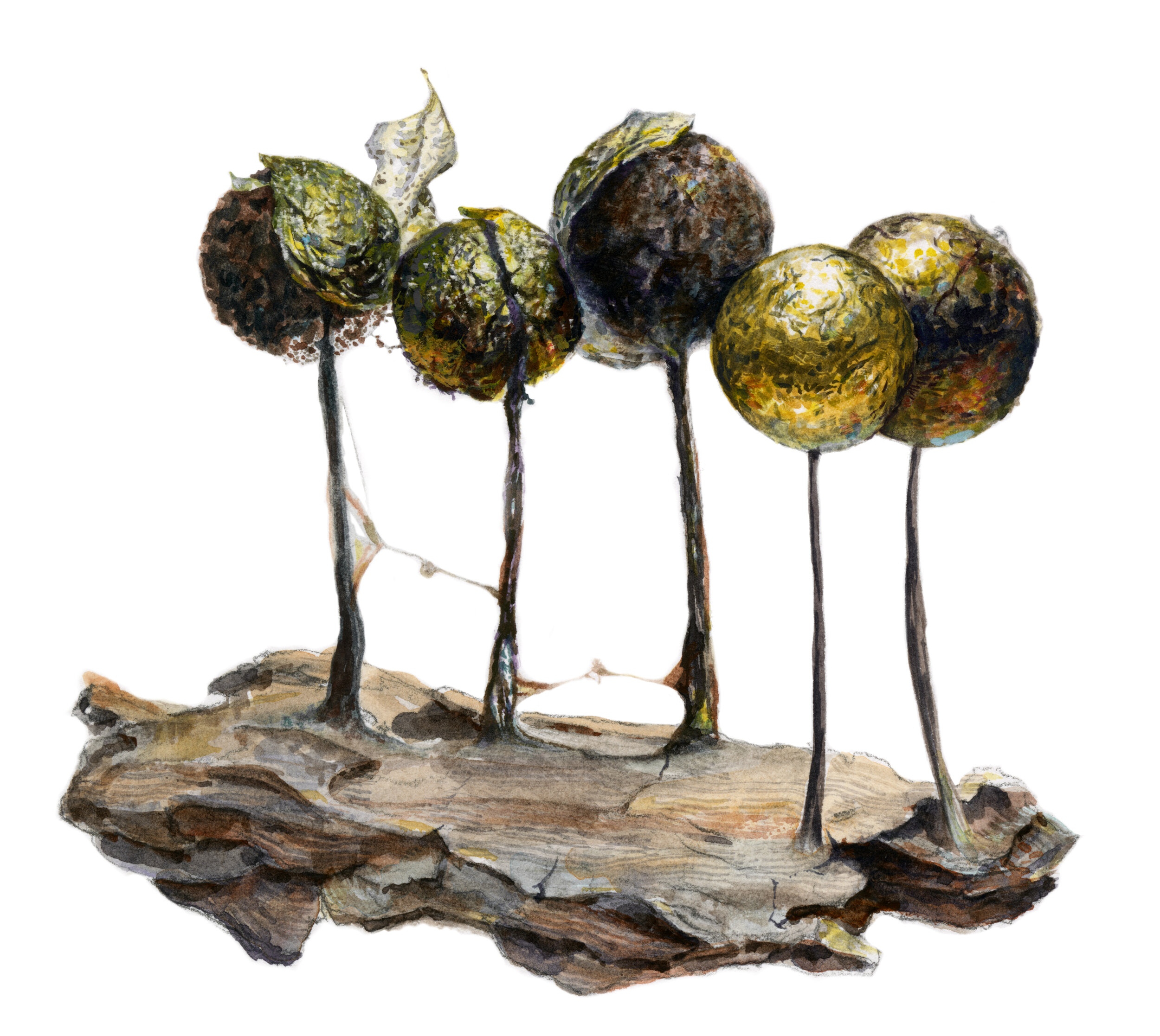
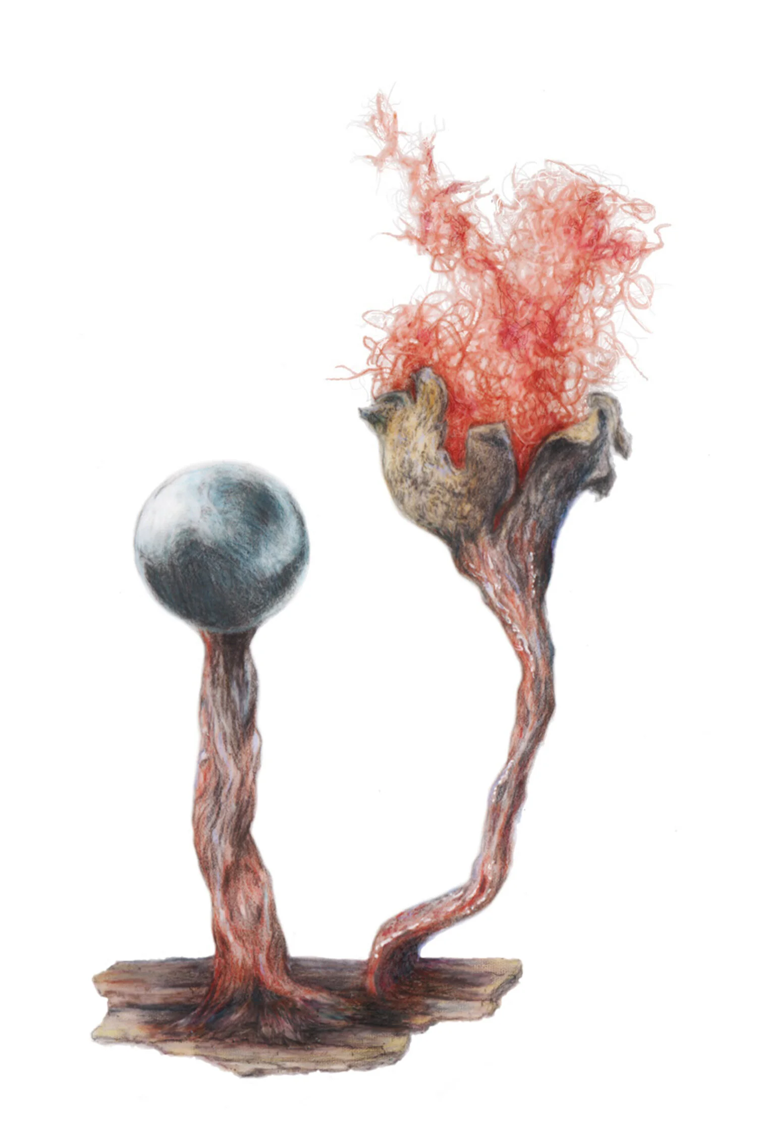
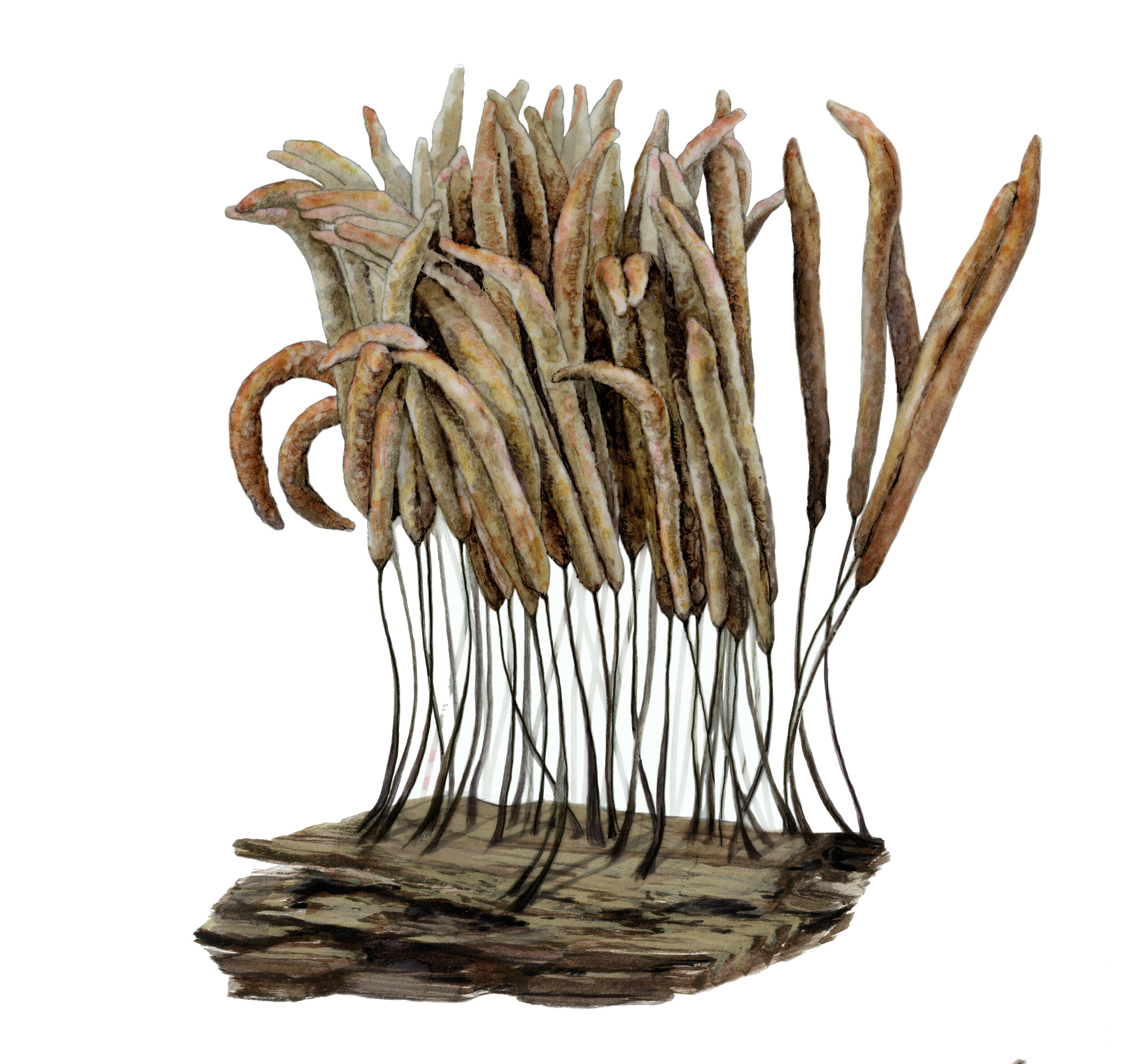
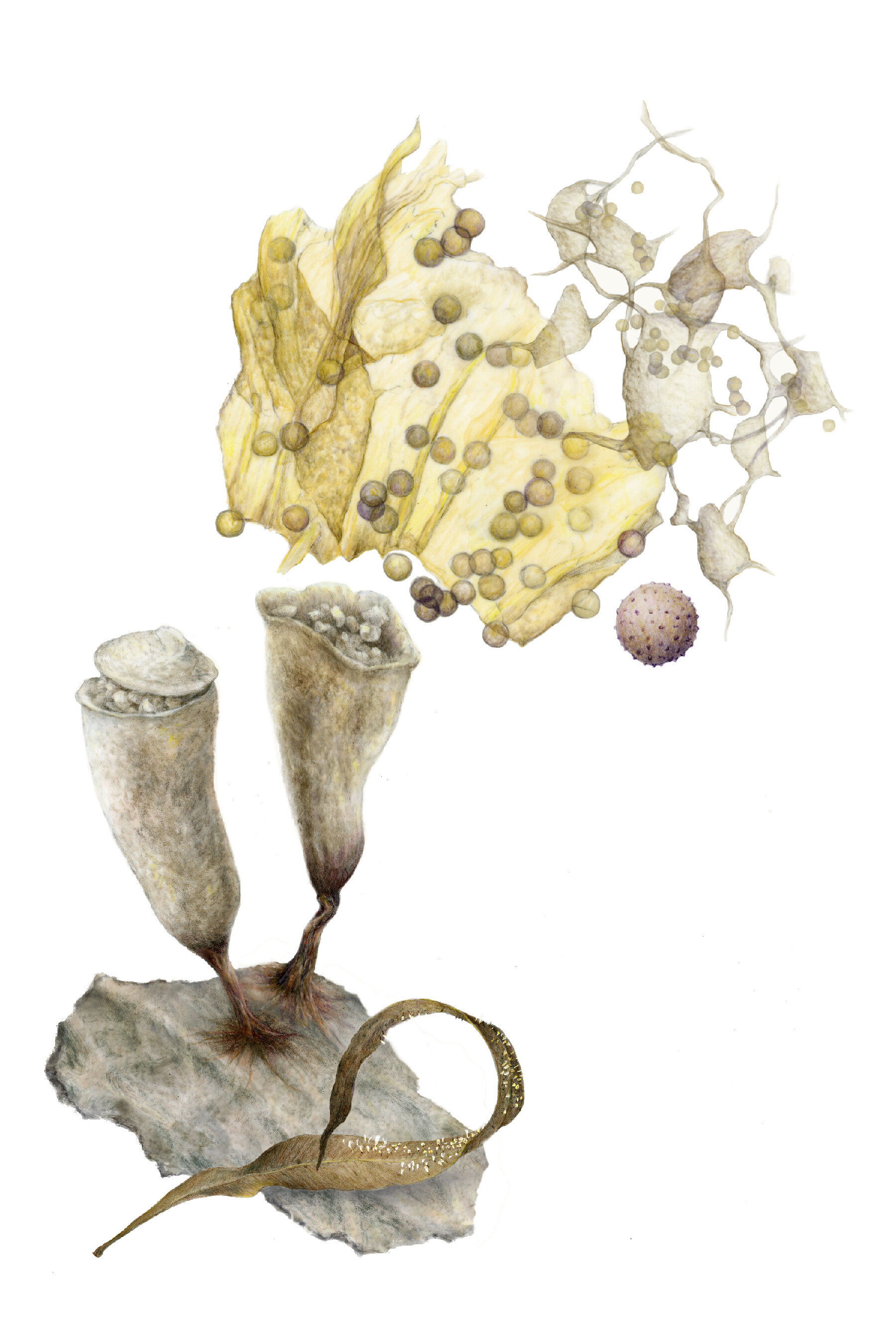
Intricate and elusive, slime molds (myxomycetes) look and “behave” unlike any other living thing. They start their lives as giant, traveling, bacteria-eating, one-celled blobs, then send up reproductive structures, each species with its own hues and ornate details. They’re common on all continents, yet most people have never seen or heard of them.
When I first “discovered” slime molds, I knew I had to find a reason to professionally illustrate them. In 2010, I learned about biologist Steve Stephenson’s research into myxomycetes’ functions in ecosystems around the world. His lab at the University of Arkansas is packed full of Petri dishes and matchbox-sized containers preserving specimens from Namibia to Tennessee. Steve invited me to illustrate species he’d found in Australia for a monograph he would work on over the next decade.
I used a microscope and mixed media to illustrate around 35 species at 40x their real size. Meanwhile I became an expert in engaging various publics in all things slime mold.
All drawings were made using specimens from Dr. Stephenson’s lab. I positioned each little specimen under a microscope and zoomed in on different “fruiting bodies” to show variations of form and stages of decay within each species.

Need more slime molds in your life?
I self-published a small, affordable book of the illustrations created for Dr. Stephenson’s more academic monograph. Check out this and other slime mold-themed goods on my sale page.

